The objective of this research is to develop a cyber-physical system
to display the inside of a patient on the skin through a 3D projector-
array and a micro camera cluster, giving the appearance of
“transparent skin” and enabling single incision surgery with the
visual benefits of open cavity surgery.
The major difficulty
in minimally invasive surgery is the loss of
natural visual perception and hand-eye coordination, which results
in a higher skill requirement, longer training and actual surgery
time. This system will give surgeons an "X-ray" vision experience,
since they see directly through the skin, and remove a spatial
bottleneck and additional scarring caused by laparoscopes.
The broad challenges being addressed in this project are reducing
the skill requirements to successfully perform an MIS (minimally
invasive surgery); reducing the invasiveness, cost, and duration of
MIS; and improving the efficiency of surgery training. The expected
outcomes of this research project will be improved dexterity for MIS
surgeons and significant economic growth in MIS and other
healthcare-related fields with numerous benefits for the nation-at-large.
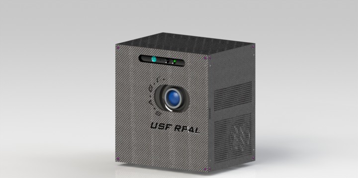
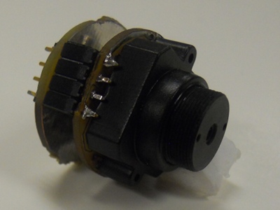
We are developing a set of micro-cameras that: occupy no space
required by surgical tools, produce no additional scarring to the
patient, and transfer wireless high-definition video images. Our
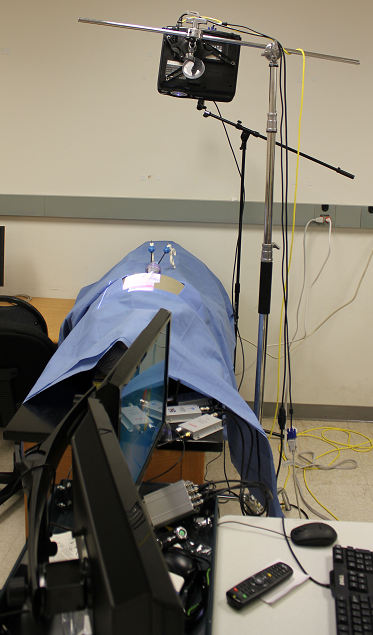
research will create a virtual view generating system to project the
panoramic 3D videos from all cameras to the right spot on the
patient’s body with geometry and color distortion compensation. A
surgeon-camera-interaction system is under-development to allow
surgeons to control viewpoint with gesture recognition and finger
tracking.
Vessel feature detection and tracking process
Vessel features detected in a Hamlyn video
Vessel features detected in another laparoscopy video
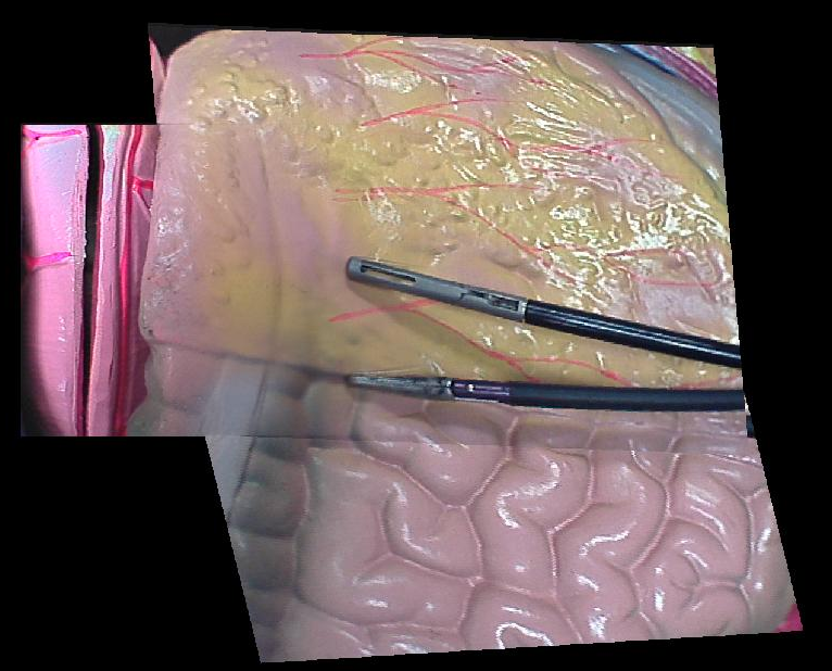
Figure 4: Mosaiced images from different cameras
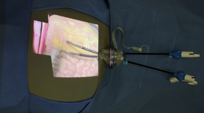
Figure 5: Projection on abdomen
This project benefits the millions of surgeries capable of being
performed through a single incision in the abdomen by providing
virtually transparent skin to surgeons who will enjoy all the visual
benefits of open-cavity surgery without all the associated risks to the
patient. The goals of this research are extremely “hands-on” and
immediately applicable to outreach activities that can excite youth,
minority students, and others about the science, medicine and
engineering careers.
Jaime Sanchez, M.D. (USF Health and Tampa General
Hospital)
Publication:
Johnson, S., Sanchez, J, French, A. and Sun, Y. (2014) Unobtrusive Augmentation of Critical Hidden Structures in Laparoscopy, MMVR, pp 1-4. (in press)
Johnson A., Sun Y. (2013) Spatial Augmented Reality on Person: Exploring the Most Personal Medium, VAMR/HCII, Part I, LNCS 8021, pp. 169-174.
Lin, B., Sun, Y., Sanchez, J., and Qian X.(2013) Vesselness Based Feature Extraction for Endoscopic Image Analysis, ISBI (in press) Data Sets
Lin, B., Johnson, A., Qian X., Sanchez, J., Sun, Y. (2013) Simultaneous Tracking, 3D Reconstruction and Deforming Point Detection for Stereoscope Guided Surgery, Augmented Reality Environments for Medical Imaging and Computer-Assisted Interventions, pp 35-44 pdf
Johnson, A. S., & Sun, Y. (2013). Exploration of spatial augmented reality on person. In IEEE Virtual Reality (VR), pp. 59-60.
Anderson, A., Lin, B., Sun Y., (2013) Virtually Transparent Epidermal Imagery (VTEI): On New Approaches To In Vivo Wireless High-Definition Video and Image Processing, IEEE Transactions on Biomedical Circuits and Systems, pp 1-9 (in Press).
Lin B., Sun Y., Qian X., (2013) Thin Plate Spline Feature Point Matching for Organ Surfaces in Minimally Invasive Surgery Imaging, SPIE Medical Imaging, pp. 1-6 (accepted, oral presentation).
Sun Y. Anderson A, Castro C, Lin B, Gitlin R (2011) Virtually Transparent Epidermal Imagery for Laparo-Endoscopic Single-Site Surgery, International Conference of the IEEE Engineering in Medicine and Biology Society (EMBC'11), pp. 2107-2110, Boston, MA, USA, August 30 - September 3, 2011. (pdf)
Any opinions, findings, and conclusions or recommendations expressed in this material are those of the author(s) and do not necessarily reflect the views of the National Science Foundation
Department of Computer Science and Engineering • 4202 E. Fowler Ave • Tampa, FL 33620 •
(813)974-7508 Created by: Emmanuel Stinson - Send comments to
estinson@mail.usf.edu
 We are developing a set of micro-cameras that: occupy no space
required by surgical tools, produce no additional scarring to the
patient, and transfer wireless high-definition video images. Our
research will create a virtual view generating system to project the
panoramic 3D videos from all cameras to the right spot on the
patient’s body with geometry and color distortion compensation. A
surgeon-camera-interaction system is under-development to allow
surgeons to control viewpoint with gesture recognition and finger
tracking.
We are developing a set of micro-cameras that: occupy no space
required by surgical tools, produce no additional scarring to the
patient, and transfer wireless high-definition video images. Our
research will create a virtual view generating system to project the
panoramic 3D videos from all cameras to the right spot on the
patient’s body with geometry and color distortion compensation. A
surgeon-camera-interaction system is under-development to allow
surgeons to control viewpoint with gesture recognition and finger
tracking.